Description
- -Monocot Stem model for biological study
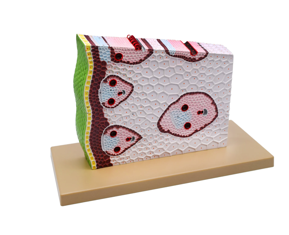
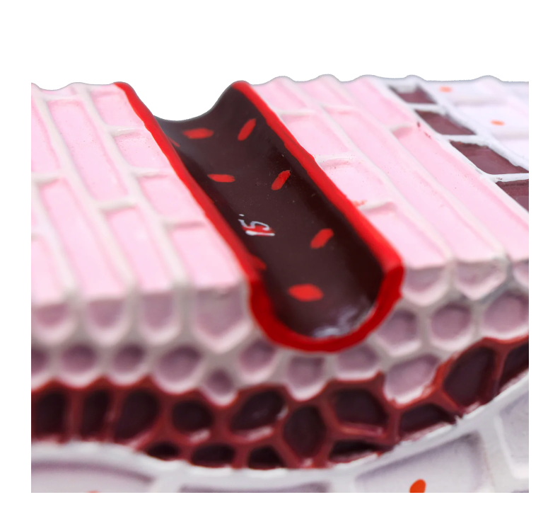
- -Greatly magnified, cross sectional view depicts structures within a monocot stem
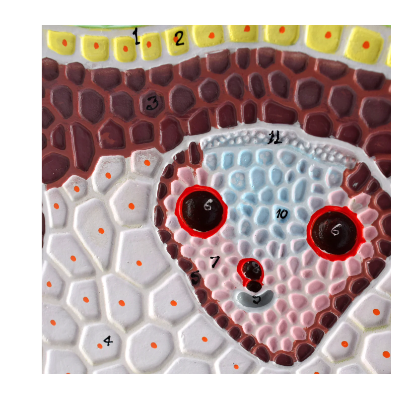
- -Numbered with key card for identification of features
- -Mounted on sturdy, ABS plastic base for display
- -Perfect for classrooms
This monocot stem model is a detailed and vibrantly colored 3D rendering that effectively shows the internal anatomy and vasculature of a monocot stem. This model of a piece of a cross-section provides an internal view of a monocot stem, and provides an excellent visual for understanding its structure and function. Key features are colored and numbered for comparison with the key that is included. This model provides a visually and kinesthetically effective method for studying the structure and function of the various structures of a monocot stem. The model is mounted on a sturdy ABS plastic base for display.
The model (not including the base) measures 12.5″ wide, 2.5″ long and 10″ tall. The base measures 18″ wide, 8″ long and 1″ high. The total height of this model measures 11″
Details Identified: cuticle, epidermal cells, hypoepidermis scleranchymatous, ground tissue parenchymatous, bundle sheath scleranchymatous, metaxylem vessels, xylem fibers, protoxylem, air cavity, phloem, protophloem, sieve tube, companion cell, sieve plate, pitted vessel, annular vessel, and spiral vessel.
Additional information
| Weight | 4 lbs |
|---|---|
| Dimensions | 11 × 18 × 8 in |



Reviews
There are no reviews yet.